
Bakers Cyst (also known as a Popliteal cyst), is a fluid filled lump located on the back of the knee on the medial side. A Bakers Cyst likely occurs with age and is most commonly associated with knee osteoarthritis, inflammatory arthritis or gout. However, a Bakers Cyst is sometimes evident in the young due to knee injury.
A Bakers cyst is located between the semimembranosus tendon and the medial head of the gastrocnemius tendon. On ultrasound, the Bakers cyst form a ‘question mark’ appearance (see Figure 1 & 2). The Bakers Cyst can extend into the calf and sometimes may leak and/or rupture causing a sudden onset of calf swelling and may mimic other conditions.
Historically, a Bakers Cyst was drained (aspiration), but this is not routine procedure now. This is because, if the underlying cause is knee osteoarthritis producing excess fluid, on draining the Bakers Cyst the fluid quickly returns because of the knee osteoarthritis! Therefore, today’s management would be to treat the knee osteoarthritis and not the Bakers Cyst. A Bakers Cyst may fluctuate in size depending on the knee activity levels. In acute knee osteoarthritis flare ups a joint injection maybe recommended.
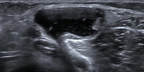
Figure 1. Bakers Cyst as demonstrated on ultrasound
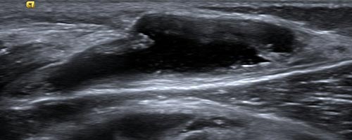
Figure 2. Bakers Cyst as demonstrated on ultrasound in longitudinal view

