

Calcific tendonitis (CT) refers to a deposit of calcium (bone) in a tendon. Commonly, CT is associated with the rotator cuff (tendons at the shoulder), but is not uncommon in the Achilles (heel), Patella (knee) and Gluteal (lateral hip) tendons.
The literature describes two types of CT; degenerative and reactive calcification. Degenerative calcification appears to be associated with ageing tendons and is likely due to calcific deposits on the tendon due to degeneration, like a frayed rope. The cause for reactive calcification is unclear, but does not appear to be due to degeneration and not always related to specific injury. The sudden onset of rotator cuff reactive CT is extremely painful causing significant arm impairment (Figure 1). Routinely, the pain is due to the calcium build up in the tendon, in combination with chemical irritation. CT is most common between the ages of 30-60 years and normally disappears in time.
Reactive tendonitis is described in the literature as having three stages:
• Pre-calcific stage: tendon changes in ways that make calcium deposits more likely to form.
• Calcific stage: calcium crystals are deposited into the tendon and are associated with the painful stage. The deposits then begin to be reabsorbed by the body and disappear.
• Post-calcific stage: tendon heals and is remodelled.
Diagnosis of CT
Asymptomatic CT is commonly found on investigations. Plain x-rays routinely identify CT, but sometimes can be missed due to the projection of the x-ray and the location of the CT. Diagnostic ultrasound (DUS) is an excellent investigation to precisely locate and measure the CT. In addition, DUS has the advantage of examining the shoulder during movement and then targeting the CT with treatment, such as an injection.
Treatment of CT
In asymptomatic cases no treatment is required. However, in symptomatic cases causing significant impairment to function and sleep deprivation there are some options available.
Routinely, analgesia and anti-inflammatory medication may control symptoms along with specialist physiotherapy. Extracorporeal shockwave therapy is non-invasive tool used to help reduce the calcium deposit and allow reabsorption. Cortisone steroid injections and/or ultrasound guided Barbotage (aspirate the calcific deposits) may be used as a more invasive procedure to reduce pain and restore function. Arthroscopic surgical decompression and excision of the calcific deposit is the last resort for recalcitrant cases.
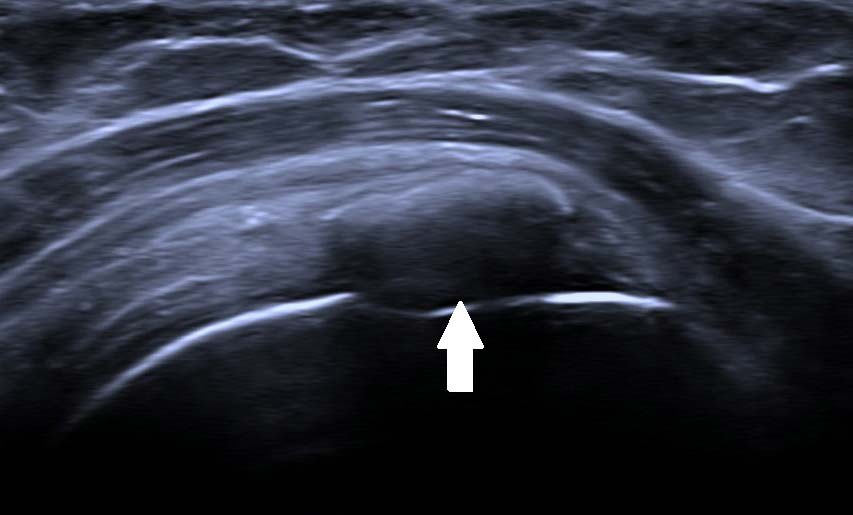
Figure 1. Supraspinatus Calcification

